Striatal Fast-Spiking Interneurons
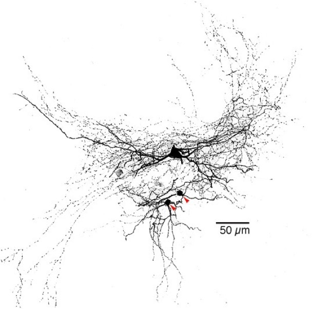
Photomicrograph of a striatal fast-spiking (FS) GABAergic interneuron biocytin-stained following whole cell recording. The two red arrowheads point to two spiny neurons that received powerful GABAergic IPSPS from the FS interneuron. Note the extremely dense axonal field of the interneuron. and the large size of the soma compared to the spiny neurons. Koós and Tepper, unpublished.
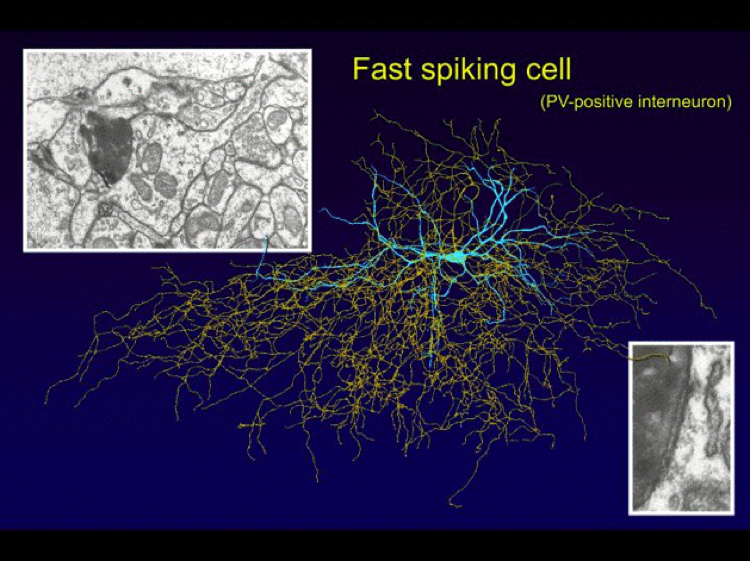
Reconstruction of a striatal FS interneuron stained with neurobiotin in vitro showing the very dense local axon collateral field (yellow). The insets show intermediate and high magnification electron micrographs of a synapse made by the interneuron onto a nearby medium spiny neuron that was simultaneously recorded. Modified from Tepper and Bolam (2004) Functional diversity and specificity of neostriatal interneurons. Curr Opin. Neurobiol., 14:684-692.
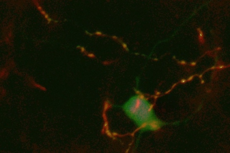
Confocal image of a biocytin filled SPN cell body (green) surrounded by local axons of a single FSI also filled with biocytin. Note the pericellular arrangement of the axons and varicosities that are presumed to be synaptic boutons. Koós and Tepper, unpublished.
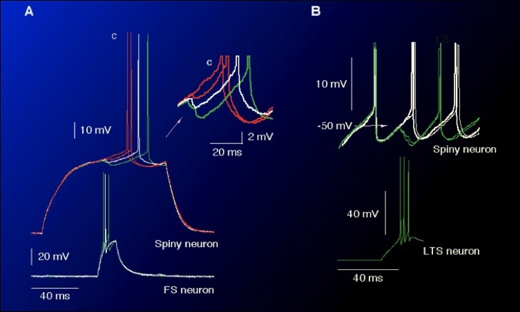
Paired whole cell recording in vitro showing interneuronal modulation of action potential generation in striatal spiny neurons. Inputs from either FS or LTS interneurons are extremely powerful and can delay or completely block evoked spiking in postsynaptic spiny neurons. See Koós and Tepper (1999) Inhibitory control of neostriatal projection neurons by GABAergic interneurons. Nature Neuroscience 2:467-472.

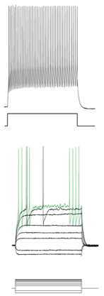
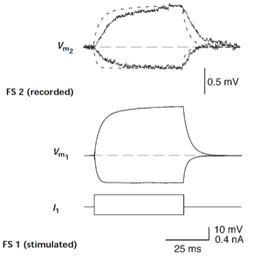
Left. Typical whole cell current clamp responses to injected current pulse in a PV+ FSI. Right. FSIs are electronicallt coupled.